Our Services
We offer a comprehensive range of dental imaging services using state-of-the-art digital technology for precise and reliable results.
Services Offered
CBCT 3D Imaging
We provide advanced Cone Beam Computed Tomography (CBCT) imaging for comprehensive diagnostic evaluation and treatment planning, including:
- Temporomandibular Joint (TMJ) Analysis
- Dental Implant Planning
- Impacted Teeth Evaluation
i-CAT FLX V17
Our office utilizes the i-CAT FLX V17, a state-of-the-art CBCT system designed to accommodate a broad range of clinical applications. As the most versatile option within the V-Series, the V17 offers a scalable field of view (FOV) of up to 23 cm x 17 cm, allowing detailed visualization of the entire oral and maxillofacial complex.
This system is widely used by orthodontists, oral and maxillofacial surgeons, and oral radiologists, and is ideal for any dental professional seeking comprehensive diagnostic imaging.
2D Imaging – Orthodontic and General Diagnostic Needs
We offer high-quality Panoramic and Cephalometric imaging to support orthodontic evaluation and a wide range of clinical applications.
Digital Intraoral Scanning
Our practice features the DEXIS IS 3800W Intraoral Scanner, delivering precise, high-resolution digital impressions for:
- Clear Aligner Therapy
- 3D Digital Study Models
- STL File Generation for Digital Workflow
Imaging Services We Offer
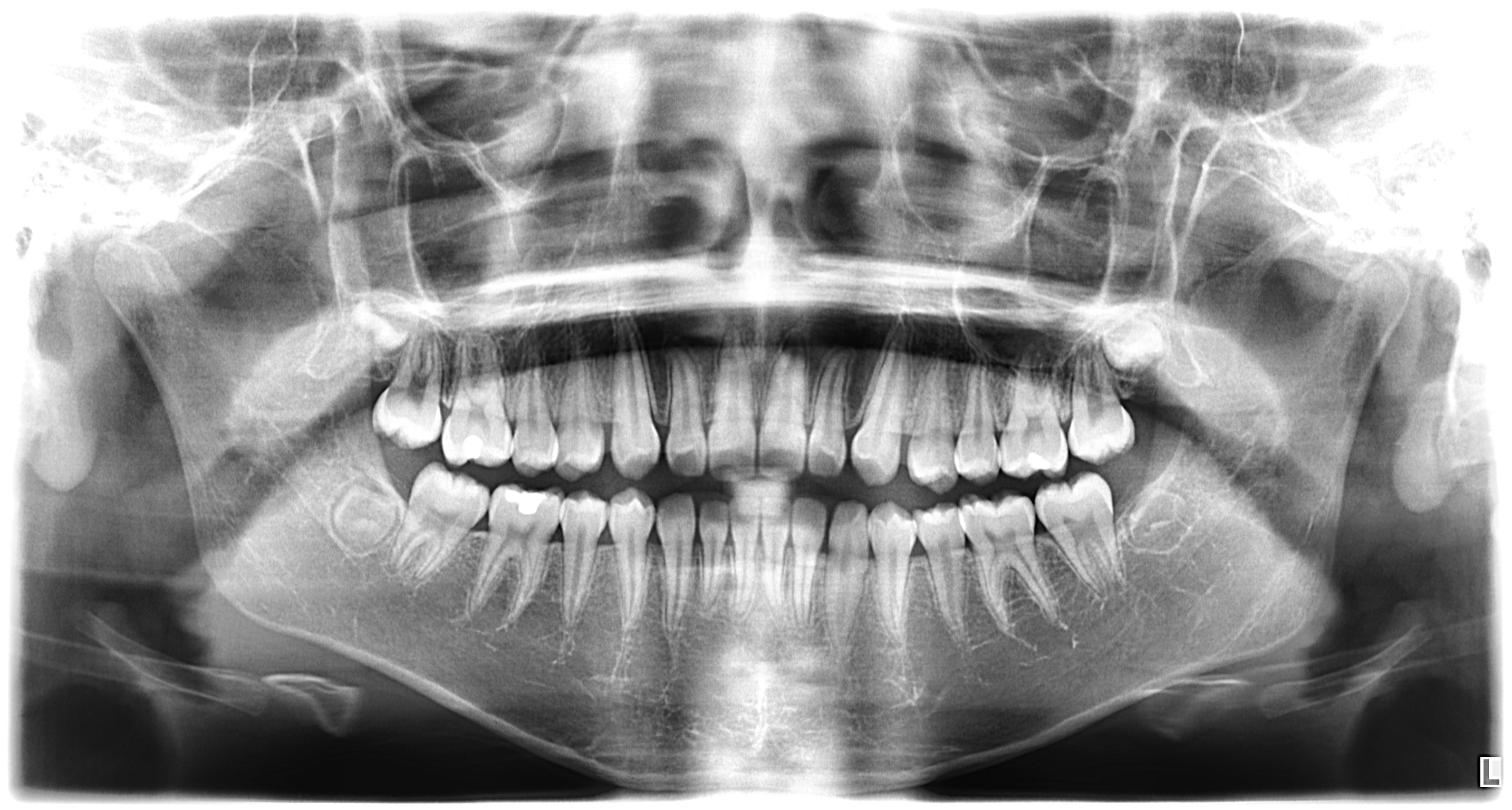
Panoramic X-Ray
A panoramic x-ray captures the entire mouth in a single image, including all teeth, upper and lower jaws, and surrounding structures. This wide-angle view is essential for evaluating overall dental health, jaw disorders, and planning treatments such as implants, braces, and extractions.
Common Uses:
- Full mouth assessment
- Implant planning
- Orthodontic evaluation
- Jaw disorder diagnosis
Example Image:
Bitewing X-Ray
Bitewing x-rays show the upper and lower back teeth in a single image. They are commonly used to detect cavities between teeth, assess the fit of dental restorations, and monitor bone loss associated with gum disease.
Common Uses:
- Cavity detection
- Crown and filling assessment
- Bone level evaluation
- Gum disease monitoring
Periapical X-Ray
A periapical x-ray captures the entire tooth from crown to root, including the surrounding bone. This detailed view helps diagnose issues at the root of the tooth, such as abscesses, cysts, and bone loss.
Common Uses:
- Root canal assessment
- Abscess detection
- Tooth fracture diagnosis
- Post-surgery follow-up
CBCT Scan
Cone Beam Computed Tomography (CBCT) provides a detailed 3D image of the teeth, bones, nerve pathways, and soft tissues. This advanced imaging technology offers unparalleled precision for complex dental procedures and diagnoses.
Common Uses:
- Implant placement planning
- TMJ evaluation
- Impacted tooth assessment
- Surgical planning
Example Images:


TMJ CBCT Scan
A TMJ CBCT scan provides highly detailed 3D imaging of the temporomandibular joints (TMJ), allowing for precise evaluation of the jaw joints, condyles, and surrounding bone structures. This specialized scan is essential for diagnosing TMJ disorders, assessing joint degeneration, and planning treatment for jaw pain and dysfunction.
Common Uses:
- TMJ disorder diagnosis
- Condyle assessment
- Joint degeneration evaluation
- Jaw pain investigation
- Pre-surgical TMJ planning
- Arthritis detection
Example Image:

Cephalometric X-Ray
A cephalometric x-ray captures a side view of the entire head, showing the relationship between teeth, jaws, and facial profile. This imaging is primarily used in orthodontic treatment planning and tracking growth patterns.
Common Uses:
- Orthodontic planning
- Growth analysis
- Surgical planning
- Facial profile assessment
Example Image:

Carpal Index
The wrist bone serves as a barometer for skeletal development. This image of the wrist may help determine the patient's developmental status and physical maturity for more confident treatment planning.
Common Uses:
- Skeletal development assessment
- Physical maturity evaluation
- Orthodontic treatment planning
- Growth prediction
PA Skull View
The posterior/anterior image displays facial symmetry, frontal and ethmoid sinuses, occipital and facial bones, and the orbital cavities.
Common Uses:
- Facial symmetry assessment
- Sinus evaluation
- Facial bone analysis
- Orbital cavity examination
Need a Different Type of Imaging?
If you need a type of dental imaging not listed above, please contact us. We are happy to discuss your specific needs and help determine the best imaging solution for your case.